

Samsun: Treatment for diabetic retinopathy includes treatment and eye follow-up planned according to the stage of diabetes-related retinal damage and the state of vision.
Diabetes can cause damage to the retinal layer, which is sensitive to light in the eye. While vision may appear normal initially, the disease can progress. The goal of treatment is to control any changes that threaten vision and to preserve existing sight; however, each patient receives a tailored treatment approach.
Samsun eye doctor : for people with diabetes, assessment should include retinal health as well as the spectacle prescription. Assoc. Prof. Muhammed Mustafa Kurt, MD : professional information is available on the home page. This text provides general patient information and does not replace an examination.
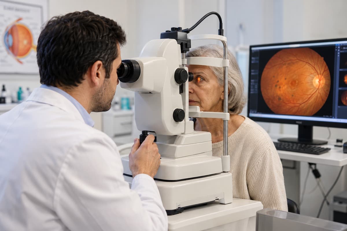
What is diabetic retinopathy?
Diabetic retinopathy is damage to the small retinal blood vessels and surrounding tissue caused by diabetes. The retina enables vision by converting incoming light into nerve signals. Vascular damage may cause fluid to leak into the retina, small haemorrhages or reduced blood flow in some areas.
A retina with reduced blood flow may release substances that stimulate the formation of new blood vessels. These developing vessels are more fragile than normal vessels. Impairment of vision depends on the extent of vascular changes, the presence of bleeding, and the condition of the macula, which is responsible for central vision.
What are the stages of diabetic retinopathy?
Diabetic retinopathy is evaluated in both non-proliferative stages (where there is no new vessel formation) and proliferative stages (where new vessels develop). The stage of the disease is determined not only by how well a person sees, but also based on the findings during a comprehensive eye examination.
The non-proliferative stage is classified as mild, moderate, and severe. In early stages, microaneurysms – small expansions – are observed in the blood vessel walls. As the condition progresses, retinal hemorrhages, leakage, and circulatory problems may increase; however, there is no new vessel formation at this stage.
In the proliferative stage, abnormal new vessels grow on the retinal surface or optic nerve head. They may bleed into the vitreous gel that fills the eye. Accompanying scar tissue can pull the retina away from its position; this is called tractional retinal detachment.
What is macular edema in diabetic retinopathy?
Diabetic macular oedema occurs when leaking blood vessels cause fluid accumulation and thickening in the macula, which is responsible for central vision. Reading, recognising faces and seeing fine detail may become difficult. Straight lines may appear curved or wavy.
Macular edema is not a separate stage of retinal disease; it can occur in different stages. Treatment decisions are made based on whether the edema is affecting the central vision, the level of vision, and findings from eye imaging. Simply detecting the edema does not necessarily mean that the same treatment will be initiated for everyone.
What causes diabetic retinopathy and who does it affect?
Diabetic retinopathy develops as a result of damage to the retinal blood vessels caused by long-term high blood sugar, and it can occur in both type 1 and type 2 diabetes. The risk increases as diabetes duration lengthens. Poor blood sugar control, as well as abnormalities in blood pressure and cholesterol levels, also contribute to the risk.
Conditions accompanying diabetes, such as kidney disease, are evaluated together with eye problems. Due to the potential for retinopathy to progress during pregnancy in individuals with a prior history of diabetes, regular eye monitoring is specifically planned. It’s not expected that all risk factors will lead to the same rate of progression.
Measuring blood sugar levels normally today does not rule out any previous retinal damage. However, regular diabetes monitoring supports healthy vision by enabling individuals to follow a personalized diet and treatment plan. Retinal examinations should not be neglected, even if metabolic control is good.
What are the symptoms of diabetic retinopathy?
Diabetic retinopathy may cause no symptoms in its early stages. As it progresses, blurred vision, floaters and dark areas in the visual field may develop. Difficulty reading, faded colours or distortion of central vision may also occur. The two eyes may not be affected equally.
These complaints are not exclusive to diabetic retinopathy. Fluctuations in blood sugar can cause temporary blurred vision, and other eye problems, such as cataracts, can also produce similar symptoms. The cause of these symptoms can be determined through examination.
Which symptoms in diabetic retinopathy require urgent evaluation?
Sudden vision loss, a sudden increase in floaters or a curtain-like shadow in vision require an eye examination on the same day. New flashes of light or a dark shadow spreading across the visual field should not be left waiting either. The absence of pain does not mean the condition is unimportant.
These symptoms can occur with problems such as bleeding inside the eye or retinal detachment; they do not establish a diagnosis on their own. Increasing eye pain, marked redness or worsening vision after treatment also require assessment without waiting for the routine follow-up.
How is diabetic retinopathy diagnosed?
The diagnosis of diabetic retinopathy is made through a dilated eye exam, examining the retina, and, if necessary, with additional imaging tests. Visual acuity and intraocular pressure are also assessed. The duration of diabetes, medications used, previous eye treatments, and recent blood sugar levels are all investigated.
The examination isn't just performed to identify the reason for a single complaint. It’s also necessary to detect and monitor vascular changes that don’t present with symptoms, and to assess whether there’s any progression. A single normal measurement or good vision cannot replace a detailed retinal evaluation.
Fundus Examination, Optical Coherence Tomography (OCT) and Retinal Angiography
Fundus examination assesses vascular damage, OCT assesses retinal thickness and fluid, and retinal angiography assesses circulation and areas of leakage. These methods provide different, complementary information. Not every patient needs all tests.
During eye examinations, hemorrhages, vascular changes, and the formation of new blood vessels are examined. Retinal photographs can allow for comparison with subsequent examinations. Given that pupil-expanding drops can cause temporary blurred vision and sensitivity to light, the transportation arrangements following the examination should be planned accordingly.
Optical coherence tomography (OCT), commonly referred to as OCT, produces cross-sectional images of the retina using light. It is particularly used to monitor the location and response to treatment of macular edema. In retinal angiography, when necessary, the passage of a dye administered intravenously through the vessels is visualized as it flows through the retinal blood vessels.
Before an angiography, any history of allergies, pregnancy, and accompanying illnesses should be reported. Whether the test is necessary and the individual’s specific potential risks will be evaluated by an ophthalmologist.
How is diabetic retinopathy treated?
Treatment for diabetic retinopathy involves monitoring, medication, laser therapy, or surgery, depending on the severity of the condition, the presence of macular edema, bleeding, and the patient’s visual acuity. In early stages, regular monitoring and good diabetes control may be sufficient. If findings indicate a threat to vision, treatments targeting the eye are considered.
The goal of treatment is not to completely eliminate existing damage in all cases, but rather to reduce the impact of the disease and preserve vision. In some patients, vision may even improve; however, if there is permanent tissue damage, recovery may be limited. The same individual may require multiple methods over time.
Intravitreal Injections for Diabetic Retinopathy
Intravitreal injections are considered particularly for central macular oedema affecting vision and in selected cases of proliferative retinopathy. Commonly used anti-VEGF medicines reduce the action of VEGF, a signal that promotes vascular leakage and abnormal vessel growth.
The procedure is performed under appropriate sterile conditions after numbing the ocular surface. Medication is delivered into the vitreous cavity. The medicine and intervals between treatments are determined from examination and OCT findings, response to previous treatment and accompanying conditions.
Steroid-containing eye treatments may also be considered in selected patients. These options should be evaluated separately with regard to eye pressure and cataracts. Not all medications are suitable for every macula edema; in some patients with good vision, an initial period of close monitoring may be preferred.
The treatment may not be fully completed with a single application. Improvement in vision does not necessarily mean that the need for continued monitoring has ended. In particular, if follow-up is discontinued in patients whose condition is being controlled with medication, the disease may reactivate.
Laser Treatment for Diabetic Retinopathy (Photocoagulation)
Laser treatment is used particularly in proliferative retinopathy to control abnormal vessels and to treat selected areas of oedema. It serves a different purpose from laser procedures used to correct a glasses prescription.
Panretinal photocoagulation applies controlled laser treatment to the peripheral retina. The aim is to reduce the stimulus for new vessel growth from tissue with reduced blood supply. Treatment may require several sessions and is sometimes combined with intraocular medication.
When dealing with macular edema, targeted laser treatment focusing on specific leakage areas may be considered in suitable patients. However, it is not typically the first option for any edema affecting central vision. The need for laser treatment is determined based on the location of the edema and previous treatments applied.
When is vitrectomy surgery necessary in diabetic retinopathy?
Vitrectomy is considered for intraocular bleeding that does not clear or scar tissue that pulls on the retina and threatens vision. The vitreous gel and, when necessary, the tissues causing traction are removed. Additional retinal treatments may be performed during surgery.
Vitrectomy, Retinal surgeries may be considered; not every patient with diabetic retinopathy needs surgery. The decision depends on the duration of bleeding, the condition of the retina and whether central vision is threatened. Anatomical repair of the retina does not necessarily mean that vision will return to its previous level.
Follow-up and important considerations after treatment for diabetic retinopathy
Following treatment for diabetic retinopathy, retinal examinations and diabetes monitoring should be conducted together. Even if bleeding or swelling subside, the condition can recur. During examinations, visual acuity, retinal findings, and OCT results (when necessary) are compared.
Follow the prescribed eye drop and care instructions; do not stop medication on your own. Return to everyday activities and driving depends on vision and the procedure performed. Head positioning, activity and travel restrictions after surgery are assessed individually.
If gas has been administered into the eye during surgery, travel by air should not be undertaken until the physician confirms that all traces of the gas have disappeared. If another procedure requiring anesthesia is planned, the medical team must be informed if gas is present in the eye. Restrictions such as ascending to high altitudes are adjusted according to the surgeon’s recommendation.
In diabetic retinopathy, blood sugar and blood pressure control are crucial.
Maintaining healthy blood sugar levels, blood pressure, and cholesterol levels is a fundamental part of managing diabetic retinopathy. These targets are determined by the physician overseeing the patient’s diabetes. Regular medication use, a suitable diet, and an exercise plan tailored to the individual’s condition all support this process.
HbA1c provides information about average blood glucose over the previous few months. Sharing results with an ophthalmologist helps place retinal findings in the context of overall health. Closer eye monitoring may be needed when treatment rapidly changes blood glucose that has been high for a long time.
This does not mean that diabetes treatment should be discontinued. Treatment adjustments should be evaluated by healthcare professionals. Even if blood sugar levels are normal, any planned eye treatments for macular edema or new vessel formation should not be automatically canceled.
How often are diabetic retinopathy screenings performed?
The frequency of monitoring is determined based on whether retinopathy is detected, the stage of the disease, and the treatment being applied. Monitoring during active eye treatment with diabetic screening is not conducted on the same schedule.
General screening guidance recommends a comprehensive eye examination at diagnosis for type 2 diabetes and within the first five years after onset for adults with type 1 diabetes. New visual symptoms require assessment without waiting for these intervals.
For some people without detected retinopathy and with good blood glucose control, a screening interval of one to two years may be considered based on previous annual assessments. If retinopathy is present, assessment is generally needed at least annually. Intervals are shorter with progression, macular oedema or active treatment.
For people with pre-existing diabetes, planning or starting a pregnancy requires reassessment of the follow-up schedule. Assessment may be arranged before pregnancy or in the first trimester, with further checks throughout pregnancy and after delivery according to findings.
What are the risks associated with treating diabetic retinopathy?
The risks associated with diabetic retinopathy treatment vary depending on the method used; potential complications include infection, changes in eye pressure, and visual impairment. The expected benefits, potential risks, and consequences of foregoing treatment are all evaluated in conjunction with examination findings.
Temporary stinging or a small bleed on the eye’s surface may occur after an intravitreal injection. Less common complications such as infection inside the eye, a retinal tear or retinal detachment require serious assessment. Corticosteroid options can raise eye pressure and accelerate cataract development.
Following laser treatment, temporary blurred vision may be observed. Specifically, night and peripheral vision may be affected, particularly with extensive laser treatments applied to the retina; there may be an increase in macular edema. These possibilities are considered alongside the risk of bleeding and vision loss, which can be caused by new vessels forming.
Following vitrectomy, there is a risk of recurrent bleeding, infection, changes in pressure, cataract formation, or retinal detachment; some patients may require additional procedures. If you experience increased pain, noticeable redness, or a new decrease in vision after the procedure, you should contact your ophthalmologist immediately.
Samsun Diabetic Retinopathy Treatment Prices – 2026
In 2026, diabetic retinopathy treatment costs in Samsun depend on the assessments, treatment method and follow-up required. The same diagnosis does not mean the same procedure or number of treatments for everyone. There is therefore no single treatment fee that applies to all patients.
The primary factors influencing pricing include examinations and imaging, the need for medication or laser treatments, surgical requirements, and the follow-up process. The fact that both eyes may require different treatments can also alter the plan. Treatment selection should be based on medical necessity, not simply on price comparisons.
Conclusion
Samsun: Treatment for diabetic retinopathy requires an approach appropriate to the disease stage, together with regular retinal follow-up and diabetes control. Macular oedema, new vessel growth and bleeding inside the eye are assessed differently; the same treatment is not suitable for every patient.
Examinations before symptoms appear help detect early changes. Treatment may preserve vision or improve it in some cases; permanent damage may not recover completely. Do not wait for a routine appointment if you experience a sudden decrease in vision, many new floaters or a curtain-like shadow.
Frequently Asked Questions
Answers about recovery, vision loss and the number of treatments in diabetic retinopathy depend on the stage of the disease and the response to treatment.
Does diabetic retinopathy completely heal?
In diabetic retinopathy, bleeding and swelling may improve; however, established, permanent retinal damage does not always fully heal. Treatment aims to control the disease and preserve vision. Even if symptoms subside, regular eye examinations continue because diabetes remains a factor.
Does diabetic retinopathy lead to blindness?
Diabetic retinopathy can lead to permanent and severe vision loss, especially in advanced stages or when treatment is delayed. However, not all patients will go blind. Early diagnosis, appropriate treatment, and regular monitoring allow for timely intervention to address any changes threatening vision.
How Many Intravitreal Injections Are Used for Diabetic Retinopathy?
There is no fixed number of intravitreal injections that applies to everyone; the need is determined by examination and OCT findings. Treatment may be needed at shorter intervals initially, with the intervals adjusted according to the response. Recurring oedema or vascular activity may require additional treatment.
Can diabetic retinopathy progress without any symptoms?
Diabetic retinopathy can progress without causing any noticeable symptoms, even when vision remains normal. Changes in the blood vessels, which often develop without pain or discomfort, may go unnoticed, particularly if they don't affect central vision. Therefore, eye examinations should be part of a diabetic management plan, not just performed when blurred vision begins.
About this article
Last updated 
Examination and individual assessmentAssoc. Prof. Muhammed Mustafa Kurt, MD
Ophthalmologist · Atakum, Samsun
- Content preparation
- Editorial information prepared using medical sources.
- Medical review
- The content update date does not indicate that a medical review has been performed by the physician.
Source: the article “Diabetic Retinopathy Treatment in Samsun” on the practice’s existing website. No additional medical references were specified in the original article.
This article is for general information and does not replace a medical examination. Diagnosis, suitability for treatment and follow-up are determined individually after an eye examination.