

Macular degeneration treatment in Samsun is planned according to the type and stage of the condition, aiming to preserve vision through examination, appropriate treatment and regular follow-up.
Age-related macular degeneration is a retinal condition that can affect daily tasks such as reading and recognising faces. Monitoring and reducing risk factors are central to dry AMD, while wet AMD may require intraocular medication. It is important not to delay assessment of new visual disturbances. 1,2
Samsun eye doctor : before an examination, patients can prepare previous eye investigation results and a list of their medicines. Assoc. Prof. Muhammed Mustafa Kurt, MD’s professional background and contact details are available on the official website.
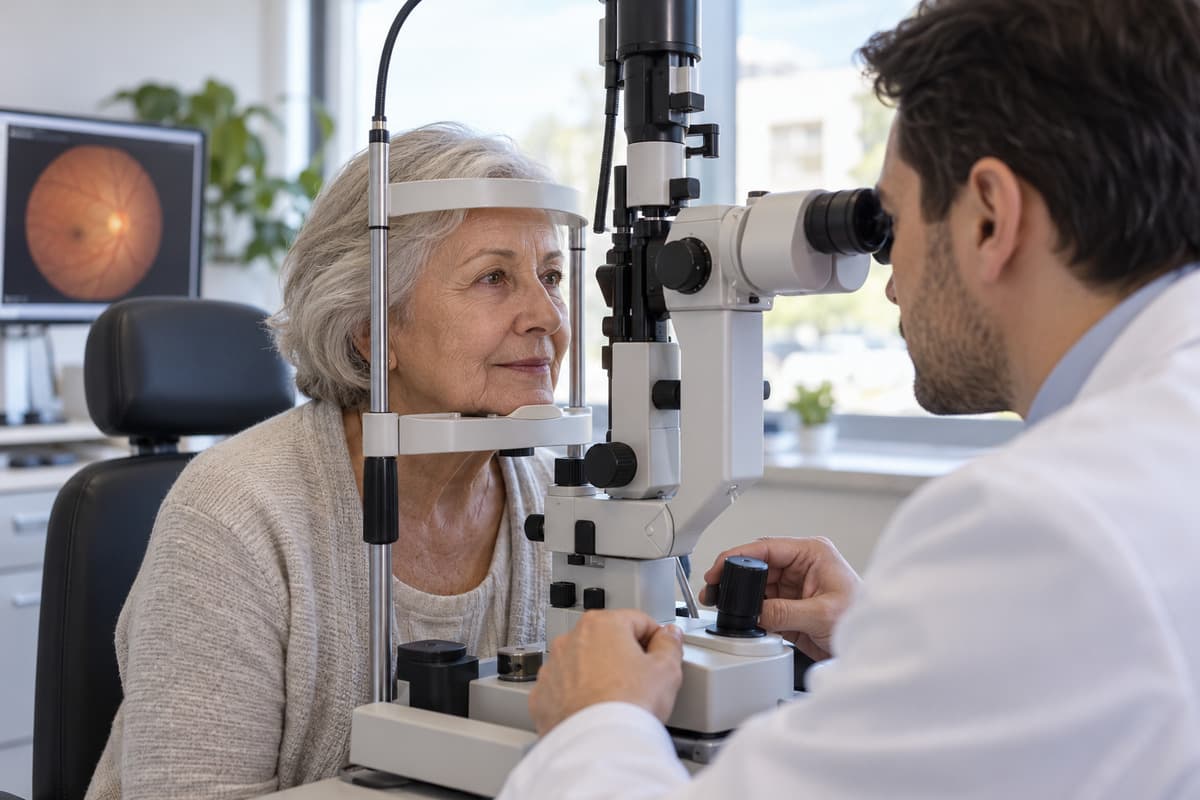
What Is Macular Degeneration?
Macular degeneration is an eye condition involving damage to the macula, the part of the retina responsible for detailed central vision.
In everyday Turkish, “sarı nokta hastalığı” usually refers to age-related macular degeneration, or AMD. The macula allows us to distinguish details in objects and faces. When this region is affected, a person may see their surroundings yet struggle to read text directly in front of them. 1
The Turkish term “sarı nokta hastalığı” does not mean a yellow spot visible on the outside of the eye. Macular holes and diabetes-related macular oedema are different conditions, even though they can affect the same region. Their treatments are not interchangeable. 3,10
What Is the Difference Between Dry and Wet Macular Degeneration?
In the dry form, cells in the macula are damaged over time; in the wet form, fluid or blood leaking from abnormal vessels affects central vision.
Dry AMD is more common and usually progresses slowly. Deposits called drusen may appear beneath the retina. In advanced disease, areas of marked cell loss may develop; this is called geographic atrophy. The dry form should not be assumed to always be mild. 4
Wet AMD can cause more rapid vision loss. Fluid accumulation and bleeding caused by leaking blood vessels disrupt macular structure. The two eyes may have different types or stages of disease. The type is established through examination and imaging, not symptoms alone. 2,14
What Causes Macular Degeneration and Who Is Affected?
Older age, family history and smoking are major risk factors for macular degeneration; the condition has no single cause.
Age-related changes in the retina and supporting tissues may combine with genetic susceptibility. The condition is more common especially after age 50. A family history of macular degeneration increases risk but does not mean the condition will definitely develop. 2
Smoking is a significant risk factor that can be modified. High blood pressure, excess weight, and cardiovascular health problems are also considered. Regular exercise, a balanced diet, and quitting smoking all contribute to overall eye health; however, there is no method to definitively prevent disease. 14
Problems with central vision that appear at a younger age can have various causes. Therefore, instead of explaining vision changes based on a person’s age or family history, it’s important to determine which part of the eye is affected. 10
What Are the Symptoms of Macular Degeneration?
Macular degeneration may cause blurred central vision, straight lines appearing wavy and a spot in the centre of the area being viewed.
The edges of the door, the window frames, or lines of text may appear wavy. Some letters may become faint while reading, and it may become difficult to discern the details of faces. Colors may appear paler than before, and reading can become difficult, especially in dim lighting. 3,4
There may be no symptoms in the early stages. When one eye sees better, a change in the other may go unnoticed. The condition is usually painless; the absence of any outwardly visible change in the eye does not mean the retina is healthy. 3
What symptoms in macular degeneration require urgent evaluation?
A sudden decrease in vision, newly distorted lines or a dark area in central vision should be assessed without delay.
When these changes occur, an ophthalmologist should be contacted the same day. Waiting because symptoms are attributed to a need for new glasses may delay treatment, particularly in wet AMD. New symptoms should be reported even if the next scheduled check is some time away. 1,11
A curtain or shadow in vision, new floaters together with flashes of light, or a painful red eye may also require urgent assessment. These can indicate serious eye problems other than macular degeneration. Sudden, marked vision loss requires attendance at the nearest emergency healthcare service. 3,10
How Is Macular Degeneration Diagnosed?
Macular degeneration is assessed using vision measurements and a retinal examination after dilating the pupil; imaging is performed when needed.
The doctor asks when the vision changes began, about family history and previous treatments. Vision is measured separately in each eye. Examination of the back of the eye assesses deposits, pigment changes, bleeding and the structure of the macula. Other eye diseases that might explain the symptoms are also investigated. 2,4
Optical Coherence Tomography (OCT) and Angiography
OCT displays retinal layers in cross-sectional images, while fundus angiography helps to evaluate vascular structure and leakage.
OCT is a diagnostic imaging technique that uses light, without employing X-rays. It helps to visualize the presence or absence of fluid within or beneath the macula, and to demonstrate any tissue changes. Comparing it with previous scans is important in evaluating treatment response and the recurrence of the disease. 10
In fluorescein angiography, the passage of dye through the eye’s blood vessels is imaged after it is injected into a vein in the arm. Other dye methods or OCT angiography without intravenous dye may be used when necessary. Not every patient needs every investigation; the choice is based on examination findings. 4,10
How Is Macular Degeneration Treated?
Treatment for macular degeneration depends on whether it is the dry or wet form, its stage and the extent of damage to the macula.
The aim of treatment is to preserve existing vision and to minimize the risk of further decline. While vision may improve in some patients, it is not always possible to completely reverse the existing damage. Treatment decisions are based not only on visual assessments, but also on a retinal examination and imaging findings. 8,13
Treatment for Dry Macular Degeneration
Follow-up, stopping smoking and vitamin-mineral supplements in suitable patients are the main approaches to dry macular degeneration.
Regular monitoring and adjustment of lifestyle habits are important in early disease. AREDS2 supplements may be considered to reduce progression risk in selected patients with intermediate disease or suitable people with advanced disease in one eye. The same supplements are not recommended for every patient with dry AMD. 6,7
For geographic atrophy in advanced dry AMD, intraocular medicines approved in some countries aim to slow enlargement of the damaged area. They do not restore lost vision. Approval, access and individual suitability in Türkiye must be assessed separately. 4,5
Low vision rehabilitation can also form part of the treatment plan when vision loss makes daily tasks difficult. Magnifying aids, appropriate lighting and screen accessibility settings support more effective use of remaining vision. 12
Treatment for Wet Macular Degeneration
Anti-VEGF medicines, which aim to reduce abnormal blood vessel growth and fluid leakage, are the main treatment for wet macular degeneration.
VEGF is a signaling protein involved in blood vessel formation. Medications that reduce the effects of this signal are administered by doctors into the eye. The goal of treatment is to control fluid and bleeding. The response can vary depending on the initial damage, the duration of the disease, and regular monitoring. 8,9
Additional methods such as photodynamic therapy may be considered in appropriately selected cases. Not every laser procedure is suitable for macular degeneration; corneal laser treatment intended to correct a spectacle prescription is not a treatment for this condition. The method is chosen according to the nature of the retinal problem. 8,10
Intravitreal Injections for Macular Degeneration
An intravitreal injection is a procedure performed by a doctor to deliver medicine into the gel-filled space in the back of the eye.
In wet AMD, the aim is for the medicine to act on abnormal blood vessels around the retina. This should not be seen as a one-off permanent solution. Treatment may need repeating when disease activity continues or returns. 8,9
How Are Intravitreal Anti-VEGF Injections Given?
Anti-VEGF treatment is administered by a physician in appropriate sterile conditions after the eye has been numbed with eye drops.
Before the procedure, the eyes and surrounding area are cleaned with an antiseptic. The medication is administered into the eye through a fine needle in a suitable location. A brief sensation of pressure or stinging may be felt; the level of discomfort varies from person to person. The preparation and post-procedure evaluation can take longer than the procedure itself. 9
The instructions for patients following a procedure and the follow-up plan are explained. Patients should inform their doctor about any medications they are taking, any allergies, and any existing health conditions before the procedure. It is important to follow the given care instructions and to consult with the unit providing the care if any new concerns arise. 9
How Many Intravitreal Injections Are Needed?
There is no number of intravitreal injections that applies to everyone; treatment intervals vary according to the medicine and the disease response.
Initially, treatment may need to be administered more frequently. Subsequently, visual measurements, examinations, and OCT findings will be evaluated together. The intervals between treatments may be extended in some patients, while others require continued frequent application. Treatment can last for months or even years. 8,13
Less fluid on OCT or better vision does not mean that treatment can be stopped without medical advice. The doctor determines follow-up and treatment decisions. If an appointment cannot be attended, notify the treating unit so the follow-up plan can be rearranged. 11,13
What Are the Risks of Intravitreal Injections?
Temporary irritation may occur after an intravitreal injection; serious eye problems can develop, although they are uncommon.
A small surface haemorrhage, stinging or temporary blurred vision may occur. Increased intraocular pressure, inflammation inside the eye, infection, bleeding and retinal detachment are more serious possibilities. Risks should be explained individually before the procedure. 9,13
Increasing pain, marked redness, reduced vision or extreme light sensitivity must not be considered normal signs of healing. Contact an ophthalmologist without waiting for the follow-up date; if one cannot be reached, seek emergency medical care. 3,9
Follow-up and Precautions in Macular Degeneration
Follow-up in macular degeneration helps detect changes in vision early and adjust treatment according to disease activity.
Recommended follow-up should continue even without symptoms. Both eyes are monitored, even if the disease is more apparent in one eye. Improving lighting, using larger print and using magnifying aids when needed can make reading easier. 11,12
If vision loss significantly affects the ability to drive or move safely, it’s important to consult with a doctor. In addition to treatment, receiving support in daily life can help reduce the impact of vision loss, but this support should not replace the medical treatment itself. 11,12
Follow-up Examinations and the Amsler Grid Test in Macular Degeneration
Follow-up frequency depends on the type of disease; the Amsler grid test can help detect new changes in central vision at home.
For the Amsler grid test, look at the centre dot on a square grid. Wear reading glasses if you use them, and assess each eye separately by covering the other in turn. Hold the grid at the distance recommended by your doctor, with adequate lighting, and keep looking at the centre dot. 15
Report new distortion of lines, missing areas, blurring or dark spots to an ophthalmologist. A normal-looking result does not rule out disease. The Amsler test does not diagnose disease or replace OCT or an examination; it is an aid to home monitoring. 15
Diet and Vitamin Supplements in Macular Degeneration
Balanced nutrition supports healthy vision; however, vitamin supplementation is recommended based on the stage of the condition and the individual’s overall health status.
A balanced diet including green leafy vegetables, a variety of fruits and an appropriate amount of fish may be preferred. No single food has been shown to cure age-related macular degeneration. Diet should not replace recommended medical treatment. 7,11
The AREDS2 approach considers specific formulations of vitamins C and E, lutein, zeaxanthin, zinc and copper. These supplements do not prevent the onset of disease or restore lost vision. Starting them for everyone at an early stage is not recommended. 6,7
For individuals who currently smoke or have smoked in the past, older formulations containing beta-carotene may not be suitable. High doses of vitamins and minerals can interact with other medications. Product selection and usage decisions should be made in consultation with a doctor, taking into account any other medications being used. 6
Macular Degeneration Treatment Costs in Samsun in 2026
The cost of macular degeneration treatment in Samsun may vary according to the scope of the examinations, investigations and treatment required.
Monitoring dry AMD and the intraocular medication needed for wet AMD involve different processes. Assessment of one or both eyes, the selected medication, imaging needs and follow-up intervals can affect the overall cost. It is inappropriate to specify a fixed number of sessions or total fee without an examination.
This page does not provide pricing information, details on discounts, or promotional offers. The scope of services and fees applied should be explained individually to each patient following a medical evaluation by the relevant healthcare facility.
Conclusion
Macular degeneration treatment in Samsun requires an approach suited to the type of disease alongside regular follow-up.
For dry AMD, the emphasis is on reducing risks and supportive treatments in suitable patients; for wet AMD, it is on controlling the disease with intraocular medication. The aim is to preserve existing vision as far as possible. New central blurring, distorted lines or sudden loss of vision require assessment without waiting for the scheduled follow-up. 1,8,11
Frequently Asked Questions
Recovery, vision loss and the need for surgery in macular degeneration are assessed in relation to the type and stage of the condition.
Can Age-Related Macular Degeneration Be Completely Cured?
There is no treatment that completely eliminates macular degeneration in every patient; the aim is to control the condition and preserve vision.
Treatment may improve vision or preserve the existing level, particularly in wet AMD. However, permanent cell loss and damage caused by scar tissue are not expected to recover completely. Follow-up should continue even if symptoms lessen. 5,8,13
Can Macular Degeneration Cause Blindness?
Macular degeneration can cause serious central vision loss; peripheral vision, however, is usually preserved.
It’s not typical to remain completely in darkness. However, reading, recognizing faces, and driving could all be significantly affected. Low vision support can help to more effectively utilize existing sight in daily tasks. 12,14
Can Dry Macular Degeneration Become Wet AMD?
Dry macular degeneration can develop into wet AMD, but this change does not occur in every patient.
Wet AMD develops when new blood vessels form and leakage occurs. Do not wait for the scheduled follow-up if you notice new distortion of straight lines or reduced central vision. Regular examinations and monitoring vision at home help identify this change. 1,15
Is Surgery Needed for Macular Degeneration?
The standard approach to macular degeneration involves follow-up and medication suited to the type of disease, rather than routine surgery.
Surgical options may be considered for certain complications or specific clinical situations. Procedures used for other conditions, such as macular holes and retinal tears, include Retinal surgeries should not be confused with the standard treatment for age-related macular degeneration. 10,16
This content is intended for general informational purposes only; it is not a recommendation for personal diagnosis or treatment. Diagnosis, treatment selection, and follow-up plans should be determined by a specialist’s examination in an eye care practice.
About this article
Last updated 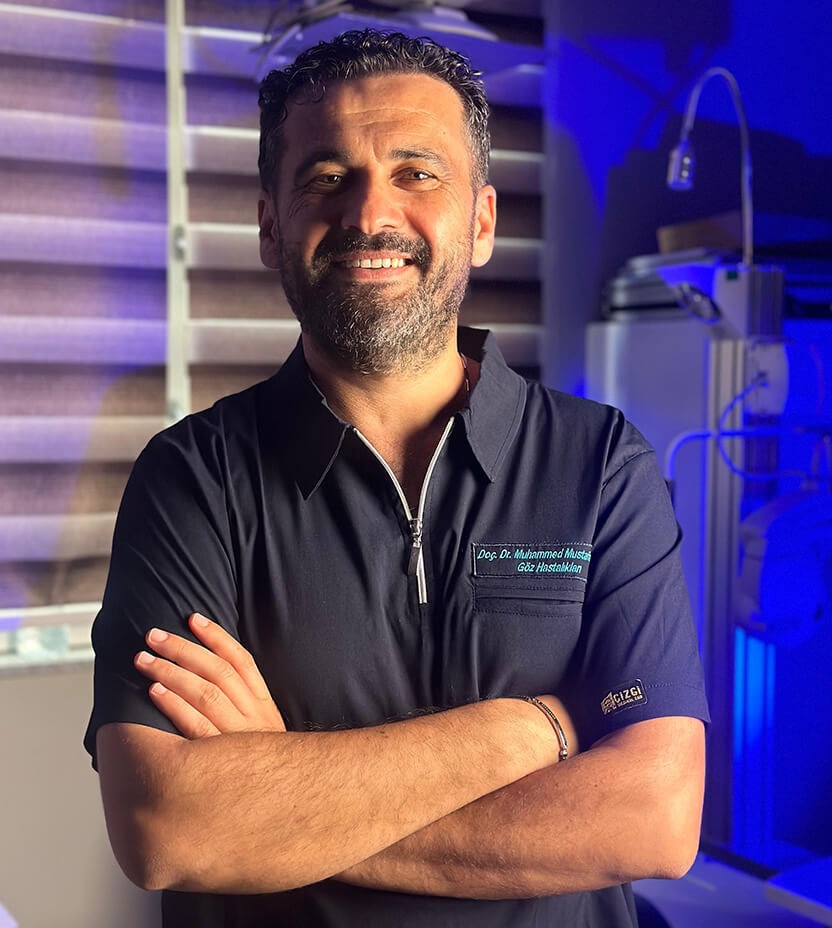
Examination and individual assessmentAssoc. Prof. Muhammed Mustafa Kurt, MD
Ophthalmologist · Atakum, Samsun
- Content preparation
- Editorial information prepared using medical sources.
- Medical review
- The content update date does not indicate that a medical review has been performed by the physician.
- Kaynak 1 · nei.nih.gov
- Kaynak 2 · asrs.org
- Kaynak 3 · nhs.uk
- Kaynak 10 · asrs.org
- Kaynak 4 · asrs.org
- Kaynak 14 · moorfields.nhs.uk
- Kaynak 11 · nhs.uk
- Kaynak 8 · nei.nih.gov
- Kaynak 13 · nei.nih.gov
- Kaynak 6 · nei.nih.gov
- Kaynak 7 · nei.nih.gov
- Kaynak 5 · fda.gov
- Kaynak 12 · nei.nih.gov
- Kaynak 9 · asrs.org
- Kaynak 15 · asrs.org
- Kaynak 16 · ema.europa.eu
This article is for general information and does not replace a medical examination. Diagnosis, suitability for treatment and follow-up are determined individually after an eye examination.