

Vitreous Haemorrhage Treatment in Samsun is planned according to the source of bleeding and retinal findings; monitoring, laser treatment or surgery may be considered.
The vitreous is a transparent gel that fills the back of the eye; hemorrhage refers to the leakage of blood into this area. The retina is the layer of the eye that perceives light. Assoc. Prof. Muhammed Mustafa Kurt, MD : assessment considers how much the blood obscures vision together with the condition of the retina.
Vitreous hemorrhage means what?
Vitreous hemorrhage is the bleeding within the vitreous gel, which is normally clear. Light may have difficulty reaching the retina; a small amount of bleeding can cause floaters, while a significant hemorrhage can lead to noticeable vision loss. The amount of bleeding alone does not indicate the severity of the condition.
The treatment of bleeding requires different goals than addressing the underlying cause that leads to bleeding. A disease requiring treatment within the retina may be found, even when vision is open. Therefore, evaluation should not be concluded simply by observing a decrease in blurriness.
Sources and risk factors for bleeding
Bleeding can develop because of fragile abnormal vessels, retinal tears or eye trauma. A condition in which diabetes damages the retina is Diabetic retinopathy : fragile new blood vessels that develop in this condition are an important source of bleeding. Retinal vein occlusions can cause similar vascular changes.
As age progresses, the vitreous gel can separate from the retina and may pull on a vessel or the surface of the retina. This separation is not the same as retinal detachment, but it can lead to the formation of tears. A history of trauma, previous eye conditions, and medications used are considered when investigating the source of bleeding.
Symptoms and need for urgent evaluation
Black spots, spider-like floaters, and hazy vision can be observed in vitreous hemorrhage. Some individuals describe a reddish curtain or a sudden decrease in vision. Bleeding can develop without pain; the absence of pain does not rule out a problem with the retina.
Sudden or rapidly increasing floaters, flashes of light, spots or shadows, and sudden vision loss should be evaluated immediately by an ophthalmologist. It is necessary to consult an eye doctor within the same day for these symptoms. Floaters do not always indicate a retinal tear; a tear and retinal detachment are investigated before deciding to monitor bleeding at home.
Examination of the retina and evaluation of bleeding.
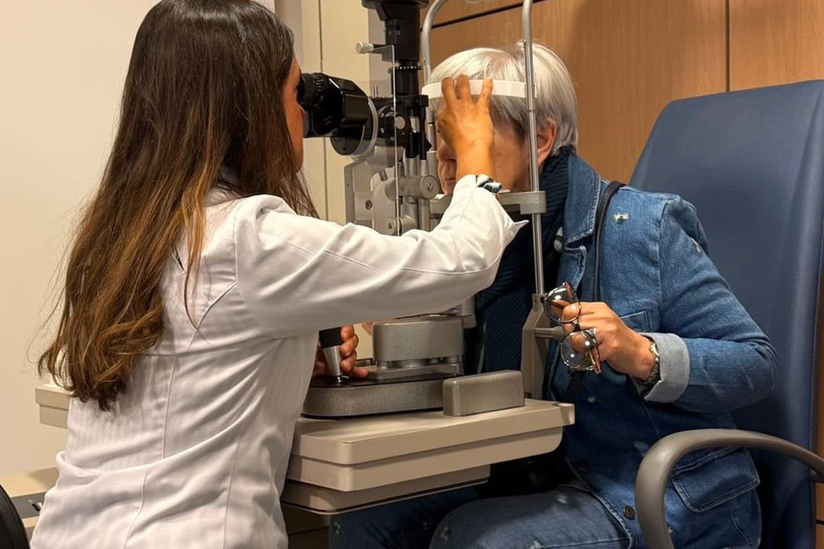
The diagnosis is determined by measuring vision and expanding the pupil to examine the retina. Bleeding onset, trauma, diabetes, and medication use are all inquired about. Samsun eye doctor : assessment also considers eye pressure, the peripheral retina and, when necessary, findings in the other eye.
When the bleeding obscures the view, ultrasound.
Ophthalmological ultrasound can assist in evaluating the posterior eye structures that may be obscured due to significant bleeding. Findings such as separation of the retina with sound waves can be investigated. Because a single examination may not completely rule out small retinal tears, follow-up examinations or further ultrasound may be required.
Consultation with a physician regarding treatment options.
Treatment depends on the cause and severity of bleeding and whether the retina is at risk. Some patients may be monitored through regular examinations. This is considered after investigating causes requiring urgent treatment; new symptoms should not wait for the scheduled follow-up.
Vision, the combined function of both eyes and recurrent bleeding also affect the decision. No single waiting period applies to every haemorrhage. Earlier intervention may be considered if the central retina is threatened or the cause of bleeding is unclear.
The place and role of laser and intraocular needle treatment.
Laser treatment and intravitreal injections may be considered to control the source of bleeding in suitable patients. For retinal tears, laser or freezing treatment may be applied around the tear. Abnormal vessels caused by diabetes may require laser treatment over wider retinal areas.
Medicines that suppress VEGF, a signal promoting abnormal vessel growth, may be injected into the eye. They provide temporary support in some patients, particularly when bleeding prevents laser treatment, but are not required for every haemorrhage. Close follow-up is important in eyes with traction on the retina.
These procedures do not mechanically drain existing blood, and they do not address all underlying causes on their own. There are risks associated with intraocular procedures, including infections and changes in pressure. The order in which these methods are used and whether they are used together are determined based on examination findings.
When Is Vitrectomy Considered?
Vitrectomy is surgical removal of blood-filled vitreous gel. It may be considered for dense bleeding that does not clear, recurrent bleeding or traction and detachment that threaten the retina. Timing depends on the cause of bleeding and the findings that threaten vision.
Gel and blood are removed through small openings; traction on the retina is relieved and laser applied when needed. Not every patient needs gas or silicone oil in the eye. Gas gradually disappears, whereas a separate procedure may be planned to remove silicone oil.
Preparation before the procedure
The medications, along with a specific anesthesia method to maintain general health and prevent pain, are considered. Any medications the patient is taking, including blood thinners, diabetes medications, and allergies, are reported to the doctor. Medication cessation or fasting decisions are made on a case-by-case basis; blood thinners are not discontinued automatically.
Temporary vision changes and any head positioning that may be needed are discussed beforehand. Support for getting home, transport to follow-up and return to work are arranged according to the procedure. Preparation and recovery schedules differ between patients.
Treatment plan, when combined with diabetes and hypertension.
Control of blood sugar and blood pressure is accompanied by eye treatment, especially in cases of retinal disease related to diabetes. When necessary, blood fats and overall health are evaluated in consultation with relevant physicians. This monitoring does not replace the necessary laser or surgical treatment for the retina.
Post-operative care and follow-up appointments.
Follow the prescribed eye drop and follow-up plan after surgery. Wash hands before using drops and do not let the bottle tip touch the eye. Avoid rubbing the eye and follow individual care instructions about contact with water.
If gas has been used, do not travel by air until it has completely disappeared. The surgical team must also provide guidance about travelling to high altitude and nitrous oxide used in anaesthesia. Decisions about head positioning, activities and driving are made individually.
Recovery of vision and potential risks
Visual recovery depends on the existing health of the retina as well as clearance of blood. Previous vascular damage or retinal disease may limit vision. Rebleeding, lens clouding (cataract), pressure changes, infection or retinal detachment may occur after surgery.
A reduction in bleeding does not necessarily indicate that the risk of recurrence has completely disappeared. During examinations, the effectiveness of the underlying condition and the state of the retina are monitored. If there is a sudden decrease in vision, severe pain, or increased redness after the procedure, immediate contact should be made with the surgical team.
Vitreous Haemorrhage Treatment Costs in Samsun in 2026
The cost varies according to the scope of billing, evaluation, and necessary treatment. It may involve examination, imaging, laser treatment, intraocular application, or surgery. Considerations include anesthesia, materials used, and the follow-up plan; a fixed price or estimated price range cannot be provided without a thorough examination.
Conclusion
Vitreous Haemorrhage Treatment in Samsun requires assessment of both the source of bleeding and the condition of the retina. The decision to monitor or intervene depends on eye findings. Sudden visual changes should be investigated promptly; necessary retinal follow-up should continue even if vision clears.
Frequently Asked Questions
Can there be bleeding within the eye, without bleeding on the white part of the eye?
Yes, bleeding within the vitreous can develop without causing redness on the white part of the eye. Bleeding in the vitreous is not the same as a red spot on the surface of the eye. The distinction cannot be made solely by looking at the appearance; if there is a change in vision, a comprehensive eye examination is required.
If bleeding has decreased, is a control examination still necessary?
Even if bleeding decreases, its cause and the retina must be checked. Areas previously obscured by blood can be examined better once the view clears. Follow-up timing depends on the initial findings; a new curtain-like shadow or reduced vision requires assessment without waiting for the appointment.
Should I myself stop taking my blood-thinning medication?
Blood-thinning medications should not be discontinued without the evaluation of a physician. The reason for taking the medication and the treatment plan should be considered together. If a specialist ophthalmologist is involved, they will coordinate any changes in the medication, and the medication will not automatically be discontinued if bleeding is observed.
Does vitreous hemorrhage develop in a person who does not have diabetes?
Even if diabetes is not present, vitreous hemorrhage can develop. Retinal tears, eye trauma, or retinal vascular diseases are among the different causes. The absence of diabetes does not eliminate the need for urgent evaluation; it is investigated through a detailed eye examination.
About this article
Last updated 
Examination and individual assessmentAssoc. Prof. Muhammed Mustafa Kurt, MD
Ophthalmologist · Atakum, Samsun
- Content preparation
- Editorial information prepared using medical sources.
- Medical review
- The content update date does not indicate that a medical review has been performed by the physician.
Source: the article “Vitreous Haemorrhage Treatment in Samsun” on the practice’s existing website. No additional medical references were specified in the original article.
This article is for general information and does not replace a medical examination. Diagnosis, suitability for treatment and follow-up are determined individually after an eye examination.