

Retinal vascular occlusions are serious eye diseases that occur when blood flow in the veins or arteries supplying the retina is partially or completely blocked. The retina is a thin layer of nerve tissue at the back of the eye that is responsible for vision. This layer needs an uninterrupted blood supply to function properly. When a blood vessel becomes blocked, the retinal tissue can no longer receive the oxygen and nutrients it needs, leading to a clinical condition that directly threatens visual function.
Retinal vascular occlusions are divided into two main groups according to the type of vessel involved: vein occlusion and artery occlusion. These two types develop through different mechanisms, produce different clinical findings and require different treatment approaches. Vein occlusions are more common and generally present with gradual vision loss, whereas artery occlusions are less common but require much more urgent intervention. In both cases, early diagnosis and timely treatment play a decisive role in preventing permanent vision loss.
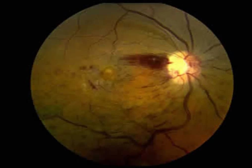
Retinal Vein Occlusion
Retinal vein occlusion is a circulatory disorder caused by blockage of the veins that collect deoxygenated blood from the retina. With vein occlusion, blood cannot drain from the retina, and the pressure that builds up in the vessels causes retinal bleeding, swelling and fluid leakage. Central vision is particularly affected when this leads to fluid accumulation in the macula. Retinal vein occlusion is the most common form of retinal vascular occlusion and usually occurs in people over the age of 50.
The severity of a vein occlusion and the area it affects depend on whether the blockage occurs in the central vein (the main vein) or a branch vein (one of the main vessel's branches). Central retinal vein occlusion affects the entire retina, whereas branch vein occlusion produces findings only in the area served by the affected vessel. Hypertension, diabetes, high intraocular pressure and blood clotting disorders are among the most common predisposing factors for vein occlusion.
Retinal Artery Occlusion
Retinal artery occlusion is an eye disease caused by sudden blockage of the arteries that carry oxygenated blood to the retina and requires urgent intervention. In artery occlusion, the oxygen supply to retinal tissue is abruptly interrupted, which can cause irreversible cell damage within minutes. The patient typically presents with sudden, painless and substantial vision loss in the affected eye. Because central retinal artery occlusion affects the whole retina, vision loss can be very severe.
Artery occlusions most often develop because of an embolus or thrombus of cardiovascular origin. Atherosclerotic plaques that break away from the carotid artery, emboli arising from heart valve disease and inflammatory vascular diseases such as giant cell arteritis are among the best-known causes. For this reason, patients diagnosed with retinal artery occlusion are referred for a systemic assessment not only for eye treatment but also to evaluate their risk of stroke and heart attack.
The Difference Between Central and Branch Occlusion
Retinal vascular occlusions are classified as central or branch occlusions according to the anatomical location of the blockage. In central occlusion, the main vessel is affected, disrupting circulation throughout the retina and causing a more severe clinical picture. Vision loss is generally more pronounced and extensive. Branch occlusion involves blockage of one branch of the main vessel, with damage occurring only in the retinal area served by that branch. Vision loss may therefore be more limited in branch occlusions.
This distinction is clinically important for both prognosis and treatment planning. Central vein occlusions carry a higher risk of macular edema and neovascularization (abnormal new blood vessel formation), and treatment is usually longer and more intensive. Spontaneous improvement is relatively more likely in branch occlusions, although treatment may be needed when the macular region is affected. Artery occlusions are similarly divided into central and branch types, with central artery occlusion carrying a much poorer visual prognosis.
Causes of Retinal Vascular Occlusion
Systemic diseases that affect blood vessel structure play a primary role in the development of retinal vascular occlusions. Atherosclerosis (hardening of the arteries) is the most fundamental predisposing factor for both artery and vein occlusions. Hypertension increases the risk of blockage by causing chronic damage to the vessel wall and narrowing the vessel lumen. Diabetes causes structural and functional changes in the retinal vascular endothelium, increasing both microvascular damage and the tendency for thrombus formation.
In addition to these systemic causes, blood clotting disorders (thrombophilias), hyperlipidemia, carotid artery stenosis, heart valve disease and inflammatory vascular diseases such as vasculitis may predispose to retinal vascular occlusion. High intraocular pressure is also considered an independent risk factor for vein occlusions. In artery occlusions, the possibility of giant cell arteritis (temporal arteritis) must be investigated, particularly in patients over the age of 70, because leaving this condition untreated may also threaten the other eye.
Risk Factors for Retinal Vascular Occlusion
The risk of retinal vascular occlusion increases significantly with age, with people over 50 forming the highest-risk group. Hypertension, diabetes, hyperlipidemia and a history of cardiovascular disease are the most common risk factors. Smoking increases the risk of occlusion by causing chronic damage to the vascular endothelium and increasing blood viscosity. Obesity and a sedentary lifestyle are also considered factors that indirectly contribute to this risk.
Glaucoma or high intraocular pressure is an independent risk factor, particularly for retinal vein occlusion. Genetic conditions that increase the tendency to clot (such as factor V Leiden mutation or protein C or S deficiency), oral contraceptive use and dehydration are also factors that should be investigated when occlusion develops in younger patients. Regular follow-up is important because patients who have had a retinal vascular occlusion in one eye may develop a similar condition in the other eye.
Symptoms of Retinal Vascular Occlusion
The most prominent symptom of retinal vascular occlusion is vision loss that develops suddenly or over a short period. In artery occlusions, vision loss usually occurs abruptly within seconds or minutes and is not accompanied by pain. In vein occlusions, vision loss may develop gradually over hours or days. Patients may describe blurred vision in the affected eye, dark areas in their field of vision or the sensation of a curtain coming down.
When the macular region is affected, prominent symptoms include marked deterioration in central vision, straight lines appearing bent or wavy (metamorphopsia) and difficulty reading. Vision loss in branch occlusions may remain limited to a particular part of the visual field, whereas central occlusions may affect the entire field. Sudden vision loss in one eye, in particular, should be regarded as a warning sign of an emergency involving retinal artery occlusion, and an ophthalmologist should be consulted without delay.
How Is Retinal Vascular Occlusion Diagnosed?
Retinal vascular occlusion is diagnosed through a comprehensive eye examination. A fundus examination directly reveals retinal hemorrhages, cotton-wool spots, venous dilation, retinal edema and changes in the macular region, providing important information about the type and extent of the occlusion. In artery occlusion, widespread retinal pallor and a characteristic cherry-red spot in the macula may be striking examination findings.
Advanced imaging methods are used to confirm the diagnosis and guide the treatment plan. OCT (optical coherence tomography) allows the presence and severity of macular edema to be assessed at the micron level. Fundus fluorescein angiography (FFA) images retinal circulation in real time, revealing the location of the occlusion, the extent of ischemic areas (areas without an adequate blood supply) and whether neovascularization has developed. Alongside these tests, blood pressure measurement and laboratory investigations such as blood glucose, lipid profile and clotting tests are integral to the assessment of underlying systemic causes.
Treatment of Retinal Vascular Occlusion
Treatment of retinal vascular occlusion is planned according to the type of blockage, the affected area and the severity of the clinical condition. Intraocular anti-VEGF injections are currently used as first-line treatment for macular edema caused by vein occlusion. These injections aim to reduce macular edema and improve visual acuity by decreasing abnormal leakage from blood vessels. Treatment is generally repeated at set intervals, and the patient's response is monitored regularly with OCT examinations.
Laser photocoagulation may be used in patients at risk of abnormal new blood vessel formation (neovascularization) in ischemic areas. Laser treatment of ischemic retinal regions aims to prevent neovascularization and related complications such as vitreous hemorrhage. In advanced cases, vitrectomy surgery may be required if vitreous hemorrhage or tractional retinal detachment develops. Treatment options for artery occlusions are more limited, and ocular massage, anterior chamber paracentesis and treatments to lower intraocular pressure during the first hours may aim to move the clot further along the vessel. Effective control of underlying systemic diseases is an integral part of treatment for all retinal vascular occlusions.
What Happens If Retinal Vascular Occlusion Is Left Untreated?
Untreated retinal vascular occlusions may lead to permanent and irreversible vision loss. In vein occlusions, chronic macular edema causes lasting damage to retinal cells over time, progressively weakening central vision. Neovascularization in ischemic retinal regions may cause vitreous hemorrhage and associated sudden vision loss. In advanced cases, abnormal vessel formation may spread to the iris and anterior chamber angle, triggering neovascular glaucoma, a painful form of high eye pressure that is difficult to treat.
In artery occlusions, the length of time retinal tissue is deprived of oxygen directly determines the visual prognosis. Untreated central artery occlusion has a high likelihood of causing severe, permanent vision loss. Moreover, artery occlusion may indicate a widespread vascular problem in the body, and the risk of life-threatening cardiovascular events such as stroke or heart attack persists if it is left untreated. For this reason, regular follow-up for both eye and systemic health is very important for patients diagnosed with retinal vascular occlusion.
Frequently Asked Questions
Can retinal vascular occlusion occur in both eyes?
Although simultaneous occlusion in both eyes is rare, people who have had an occlusion in one eye are at risk of developing a similar condition in the other. This risk is particularly increased when underlying systemic diseases are not adequately controlled. Regular follow-up of both eyes is therefore recommended.
Does vision recover completely after retinal vascular occlusion?
Visual recovery varies according to the type of occlusion, the affected area, the timing of treatment and the patient's general health. Appropriate treatment may produce a meaningful improvement in vision in branch vein occlusions, whereas the prognosis is generally poorer in central artery occlusions. Starting treatment early has a positive effect on visual outcomes.
Can retinal vascular occlusion develop at a young age?
Although more common at older ages, retinal vascular occlusion can also develop in younger people. In young patients, it is particularly important to investigate underlying causes such as clotting disorders, autoimmune diseases, oral contraceptive use and vasculitis.
About this article
Last updated 
Examination and individual assessmentAssoc. Prof. Muhammed Mustafa Kurt, MD
Ophthalmologist · Atakum, Samsun
- Content preparation
- Adapted from an information article on the practice’s existing website.
- Medical review
- The content update date does not indicate that a medical review has been performed by the physician.
Source: the article “Retinal Vascular Occlusions” on the practice’s existing website. No additional medical references were specified in the original article.
This article is for general information and does not replace a medical examination. Diagnosis, suitability for treatment and follow-up are determined individually after an eye examination.