

Keratoconus Treatment in Samsun aims to support vision and reduce disease progression according to the thinning and shape changes in the cornea.
In keratoconus, the transparent layer at the front of the eye may gradually become thinner and irregular in shape. Treatment need is not determined by the glasses prescription alone. Assoc. Prof. Muhammed Mustafa Kurt, MD : examination can assess corneal maps, visual acuity and changes over time together.
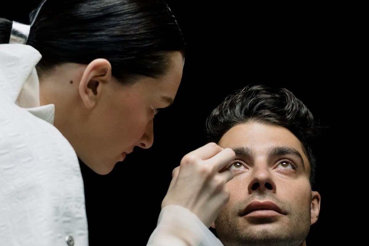
How does corneal shape affect vision?
The cornea is the clear tissue that helps focus light into the eye. Thinning in keratoconus and forward bulging can cause irregular refraction of light. Blurring, ghost images or light scatter may occur alongside myopia and irregular astigmatism.
The two eyes may not be affected to the same degree. Frequent changes in spectacle prescription or inability to achieve adequate clarity with spectacles may require investigation. Because these complaints can arise from other reasons, keratoconus cannot be diagnosed solely based on the symptoms.
Corneal mapping and assessment of progression
Diagnosis combines vision testing, assessment of refractive error and examination of the cornea. Topography or tomography provides detail about corneal shape and thickness. The findings should be interpreted alongside vision achieved with glasses and previous records.
Samsun eye doctor : bring earlier corneal maps and contact lens details to the assessment. Lenses can affect corneal measurements; ask the team how long to stop wearing them before measurement, according to lens type. Progression is not determined from one variable or the person’s description of blur alone.
Glasses and specialist contact lenses
In cases of early or milder conditions, spectacles can provide sufficient visual support. When corneal irregularities are present, options such as hard gas permeable, hybrid, or scleral lenses may be considered. Lens selection depends on the corneal structure, overall eye health, and the individual’s usage conditions.
Appropriate lenses may help to support vision by reducing the optical effect of irregular lens surfaces; however, this does not mean it will stop the progression of the disease. The initial lens may not be comfortable or suitable for every individual. Trial, fit evaluation, and follow-up are necessary; if discomfort persists, lens use should not be continued, and consultation with the lens team should occur.
What is the purpose of cross-linking?
The aim of treatment for corneal cross-linking is to reduce the progression of keratoconus in suitable eyes. Corneal tissue strength is supported using riboflavin and controlled ultraviolet light. The decision to proceed with the treatment is made based on progression findings, age, thickness, and other corneal characteristics.
Cross-linking does not directly eliminate the need for glasses. Glasses or specialised lenses may still be needed, and visual changes differ between people. Thin corneas or certain surface problems require separate suitability assessment. The same protocol is not used for every keratoconus diagnosis.
Recovery and follow-up after the procedure
Following cross-linking, temporary pain, light sensitivity, and visual fluctuations may occur. Eye drops supporting surface healing, and protective lenses may be used in some treatment methods. The duration of their use is determined according to the treatment method and examination findings.
It is important not to rub the eye and to follow the given treatment plan. It is not appropriate for the patient to attempt to put the protective lens back on themselves if it falls off. An increase in pain, redness, discharge, or a noticeable decrease in vision may indicate a problem such as an infection, and should not be expected to resolve on its own.
Intracorneal rings and transplant options
Corneal ring segments can be evaluated in selected patients in order to reshape the cornea. Lens adaptation or visual quality may improve in some individuals. These rings are not suitable for all patients and do not carry the same objective as the cross-linking procedure; progression monitoring continues.
Advanced irregularities, scar formation, or inadequate vision due to other methods may lead to corneal transplantation. The methods considered depend on the depth of the damage: partial or full-thickness grafts can be evaluated. Risks such as infection, tissue rejection, astigmatism, and prolonged healing must be discussed; optical support may be required after the transplant.
Eye rubbing, allergy and lens care
Rubbing the eyes may be associated with the development or progression of keratoconus. Instead of attempting to relieve itching by constantly rubbing, an evaluation of allergies and eye surface problems should be performed. Appropriate treatment can help reduce itching and prevent the habit of rubbing.
Learn the correct cleaning and storage plan for your lens type. Do not clean lenses with tap water or saliva, or sleep in them unless specifically permitted. Remove lenses if pain or redness develops and obtain an eye assessment before wearing them again. Lens case care is also part of safe use.
Sudden blurring and long-term monitoring
In keratoconus, a sudden and noticeable decrease in vision should be evaluated without delay. In advanced cases, fluid accumulation in the cornea can lead to edema, resulting in sudden blurred vision and, sometimes, pain. Trying new glasses or drops at home should not delay the evaluation of this condition.
Regular monitoring involves comparing vision, lens fit, corneal thickness and shape changes. The monitoring interval varies according to age, progression risk, and previous treatments. Procedures such as LASIK, which remove corneal tissue, are generally not suitable in keratoconus; surgical decisions are made considering corneal characteristics.
Keratoconus Treatment Costs in Samsun in 2026
In the evaluation for keratoconus, the examination, corneal measurements, and the scope of the chosen method are important. Special lens fitting, cross-linking, rings, and grafts are separate processes. It should be explained which tests are required; a standard treatment package and a fixed fee should not be recommended without a diagnosis or evidence of progression.
Conclusion
Keratoconus Treatment in Samsun is planned according to corneal structure and changes in the condition. Optical options that support vision and procedures intended to reduce progression have different aims. Appropriate measurements, individual method selection and long-term follow-up help meet daily visual needs.
Frequently Asked Questions
Can I stop wearing glasses after cross-linking?
This procedure primarily aims to slow the progression. It may require spectacles or lens; individual visual responses are evaluated during testing.
Do rigid contact lenses stop disease progression?
Lens vision support is used. Progression control requires a separate evaluation; seeing well does not eliminate the need for follow-up.
Does everyone diagnosed with keratoconus need a corneal transplant?
No. Many people can be followed with glasses, contact lenses or other appropriate approaches. Transplantation is considered particularly in more advanced and selected cases.
Can I wait if one eye suddenly becomes very blurred?
Sudden, intense blurred vision may be associated with hydrops or other serious problems. An immediate eye examination should be obtained.
About this article
Last updated 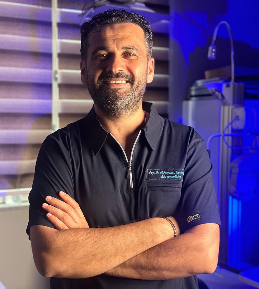
Examination and individual assessmentAssoc. Prof. Muhammed Mustafa Kurt, MD
Ophthalmologist · Atakum, Samsun
- Content preparation
- Editorial information prepared using medical sources.
- Medical review
- The content update date does not indicate that a medical review has been performed by the physician.
Source: the article “Keratoconus Treatment in Samsun” on the practice’s existing website. No additional medical references were specified in the original article.
This article is for general information and does not replace a medical examination. Diagnosis, suitability for treatment and follow-up are determined individually after an eye examination.