

Retinitis pigmentosa is an inherited eye disease characterized by progressive degeneration of the photoreceptor cells in the retina. The retina is the light-sensitive nerve layer at the back of the eye and contains two main types of photoreceptor cells responsible for vision. Rod cells are responsible for vision in the dark and peripheral vision, while cone cells are responsible for central vision and color perception. In retinitis pigmentosa, rod cells are affected first, so the disease typically presents with impaired night vision and narrowing of the peripheral visual field.
The disease usually begins in childhood or adolescence and progresses slowly but continuously over the years. In advanced stages, central vision loss may also develop as cone cells become affected. Retinitis pigmentosa is a rare disease affecting approximately one in every 4000 people worldwide, but it is the most common form of inherited retinal dystrophy. The course of the disease can vary greatly between individuals. Some patients retain a functional level of vision into middle age, while others experience earlier and more rapid progression of vision loss. Although there is currently no definitive treatment that completely stops the disease, research in areas such as gene therapy and retinal implants offers promising developments.
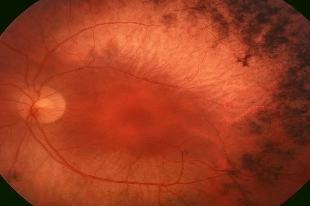
What Causes Retinitis Pigmentosa?
Retinitis pigmentosa is an inherited disease caused by genetic mutations. To date, hundreds of mutations identified in more than 80 different genes have been associated with retinitis pigmentosa. These genes largely encode proteins involved in the structure, function and metabolism of photoreceptor cells. As a result of these mutations, photoreceptor cells become unable to maintain their normal functions and are gradually lost through programmed cell death (apoptosis). This cell loss begins at the periphery of the retina and progresses toward the center.
The disease can be passed on through three different inheritance patterns. In the autosomal dominant form, a single mutated gene from one parent is sufficient for the disease to develop, and this form generally has a milder course. In the autosomal recessive form, a mutated gene must be inherited from both parents, who may be healthy carriers. The X-linked recessive form predominantly affects males and generally causes earlier onset and a more severe course. In some cases, the disease may arise through de novo (new) mutations without a family history. Because of genetic heterogeneity, the severity and rate of progression can differ even within the same family.
Symptoms of Retinitis Pigmentosa
The first and earliest symptom of retinitis pigmentosa is night blindness (nyctalopia). Because rod cells are primarily affected, the patient has marked difficulty seeing in dark or dim environments. Typical early complaints include difficulty walking outside in the evening, the eyes taking a very long time to adapt on entering a dark room and a significant decline in visual quality when driving at night. These symptoms often begin to be noticed in childhood or adolescence, but the patient may regard them as normal for their age.
As the disease progresses, the peripheral visual field gradually narrows. The patient first becomes unable to notice objects at the edges of the visual field, which then narrows toward the center over time, producing a condition known as tunnel vision. At this stage, the patient can see the point they are looking at directly but cannot perceive objects around it. Serious difficulties arise when going up and down stairs, walking in crowded places and noticing obstacles. In advanced stages, degeneration of cone cells may also lead to deterioration in central vision, changes in color perception and difficulty reading.
Stages of Retinitis Pigmentosa
Although retinitis pigmentosa does not have a clearly defined clinical staging classification, its course can generally be described as early, intermediate and advanced. In the early stage, degeneration of rod cells has begun, but vision loss is still limited. The patient may notice difficulty with night vision, and characteristic pigment deposits called bone spicules begin to appear in the peripheral retina on fundus examination. Central vision is usually preserved at this stage, and the patient can remain largely independent in daily life.
In the intermediate stage, the peripheral visual field has narrowed considerably and tunnel vision is developing. Retinal vessels have become attenuated, the optic disc has taken on a waxy pale appearance and pigment deposits have spread across larger areas of the retina. The patient experiences increasing difficulty with daily activities and may need assistive devices. In the advanced stage, both peripheral and central vision are severely affected. As cone cells also degenerate, central visual acuity decreases, color discrimination becomes difficult and some patients may retain only light perception. The time taken to move between stages varies greatly between individuals and may range from a few years to several decades.
How Is Retinitis Pigmentosa Diagnosed?
Retinitis pigmentosa is diagnosed by assessing clinical examination findings together with functional tests and imaging methods. A dilated fundus examination looks for the disease's characteristic triad: bone-spicule-like pigment deposits in the peripheral retina, attenuation of the retinal arteries and a waxy yellow pallor of the optic disc. Although these findings become more evident in advanced stages, the fundus examination may appear normal in early stages, and functional tests are needed to support the diagnosis.
OCT (optical coherence tomography) evaluates the thickness of the retinal layers and the condition of the photoreceptor layer at the micron level, revealing the extent of degeneration and the degree to which the central retinal structure is preserved. Fundus autofluorescence imaging is useful for assessing disease activity and patterns of progression by showing changes in retinal pigment epithelium metabolism. Genetic tests are used to identify the responsible gene mutation and determine the inheritance pattern. Genetic diagnosis is increasingly important in assessing a patient's suitability for new treatment options such as gene therapy.
Electroretinogram (ERG) Test
The electroretinogram is considered the most valuable functional test for diagnosing retinitis pigmentosa. It measures the retina's electrical response to a light stimulus and objectively evaluates photoreceptor cell function. During the test, a special contact lens electrode is placed on the patient's corneal surface, and electrical signals originating from the retina in response to light stimuli under different lighting conditions are recorded. Scotopic recordings after dark adaptation assess rod cell function, while photopic recordings in light conditions assess cone cell function separately.
In retinitis pigmentosa, ERG responses are characteristically reduced or completely absent. A marked reduction in rod cell responses on ERG may be detected even during early stages when the fundus examination does not yet show clear findings. This makes ERG extremely valuable for early diagnosis of retinitis pigmentosa. ERG is also an important diagnostic tool for monitoring disease progression, evaluating the response to treatment and distinguishing retinitis pigmentosa from other retinal degenerations. Multifocal ERG separately maps function in different retinal regions, allowing assessment of how well central retinal function is preserved.
Visual Field Test
Visual field testing (perimetry) is a fundamental diagnostic tool for assessing the extent and progression of peripheral vision loss in patients with retinitis pigmentosa. During the test, the patient focuses on a point while their ability to detect light stimuli appearing in different parts of the visual field is recorded. Retinitis pigmentosa typically produces a ring-shaped area of visual field loss (ring scotoma) that begins in the mid-periphery. As the disease progresses, this area of loss expands, progressively narrowing peripheral vision and eventually moving toward the center to produce tunnel vision.
Visual field testing is repeated regularly to determine the stage of the disease, monitor the rate of progression and evaluate the response to treatment. As a dynamic test, Goldmann perimetry offers advantages for assessing large peripheral areas, while computerized (automated) perimetry provides more standardized and reproducible measurements. Visual field test results are also an important reference for evaluating a patient's everyday functioning. Visual field data directly contribute to clinical decisions about eligibility for a driving license, occupational capacity and rehabilitation needs.
Treatment of Retinitis Pigmentosa
There is currently no definitive treatment that completely stops or cures retinitis pigmentosa. However, the main goals of disease management are to slow photoreceptor degeneration, preserve existing visual function and improve quality of life. Vitamin A palmitate supplementation has been reported to slow the rate of decline in ERG responses in some patients with retinitis pigmentosa, but this treatment is not suitable for every patient and must be administered under medical supervision because of the potential side effects of high-dose vitamin A.
Gene therapy is the most promising area of development in the treatment of retinitis pigmentosa. An approved gene therapy product is available for retinal dystrophy caused by an RPE65 gene mutation; it is administered by injecting a functional copy of the gene beneath the retina in patients with the appropriate gene mutation. This success has accelerated research into treatments for other gene mutations. Retinal implants (bionic eyes) are technological solutions developed to provide some visual function for patients with advanced disease. Optogenetic therapy and stem cell applications are other approaches under investigation. Supportive approaches such as low vision rehabilitation, magnifying lens systems, screen readers and orientation training also make an important contribution to maintaining patients' independence in daily life.
Eye Diseases Associated with Retinitis Pigmentosa
Retinitis pigmentosa may occur as an isolated retinal disease or alongside additional eye conditions in some patients. The most common associated eye disease is posterior subcapsular cataract. A substantial proportion of patients with retinitis pigmentosa develop cataracts in the posterior capsular region, even at a young age. This type of cataract may affect vision early but can be treated successfully with cataract surgery, allowing better assessment of existing retinal function.
Cystoid macular edema is another common complication in patients with retinitis pigmentosa and adversely affects central vision. This condition, which is readily identified on OCT, can be treated with topical or oral carbonic anhydrase inhibitors. Myopia, vitreous opacities and glaucoma may also accompany retinitis pigmentosa in some patients. Retinitis pigmentosa may additionally occur as part of certain syndromes. Usher syndrome is the best-known syndrome in which retinitis pigmentosa and hearing loss occur together. Bardet-Biedl syndrome is a rare multisystem condition that includes findings such as retinitis pigmentosa, obesity, kidney abnormalities and polydactyly. Hearing assessment and systemic screening when needed are therefore recommended for patients diagnosed with retinitis pigmentosa.
Frequently Asked Questions
Does retinitis pigmentosa cause blindness?
The course of retinitis pigmentosa varies greatly between individuals. Some patients retain functional central vision into old age, while others develop more noticeable peripheral and central vision loss earlier. Complete blindness is not an inevitable outcome in every case, but vision may decline to the level of light perception in advanced stages.
Can retinitis pigmentosa be passed on to children?
Because retinitis pigmentosa is a genetic disease, it carries a risk of being passed on to children. The likelihood of transmission varies according to the inheritance pattern.
Can patients with retinitis pigmentosa drive?
Fitness to drive depends on the patient's current visual acuity and the extent of their visual field. Safe driving may not be possible when the peripheral visual field falls below a certain threshold. Visual field test results are the main reference for this assessment, and the decision is based on the individual's current examination findings.
About this article
Last updated 
Examination and individual assessmentAssoc. Prof. Muhammed Mustafa Kurt, MD
Ophthalmologist · Atakum, Samsun
- Content preparation
- Adapted from an information article on the practice’s existing website.
- Medical review
- The content update date does not indicate that a medical review has been performed by the physician.
Source: the article “Retinitis Pigmentosa” on the practice’s existing website. No additional medical references were specified in the original article.
This article is for general information and does not replace a medical examination. Diagnosis, suitability for treatment and follow-up are determined individually after an eye examination.