

Retina care in Samsun addresses problems of the eye's light-sensitive layer through symptoms, detailed investigations, medical or surgical treatment and long-term follow-up.
Retinal conditions may cause similar symptoms, yet their treatment goals and urgency can differ. The macula is the part of the retina that contributes to detailed central vision. During an assessment with Assoc. Prof. Muhammed Mustafa Kurt, MD, the location of the condition, its effect on vision and follow-up needs can be discussed together.
The scope of retinal and macular conditions
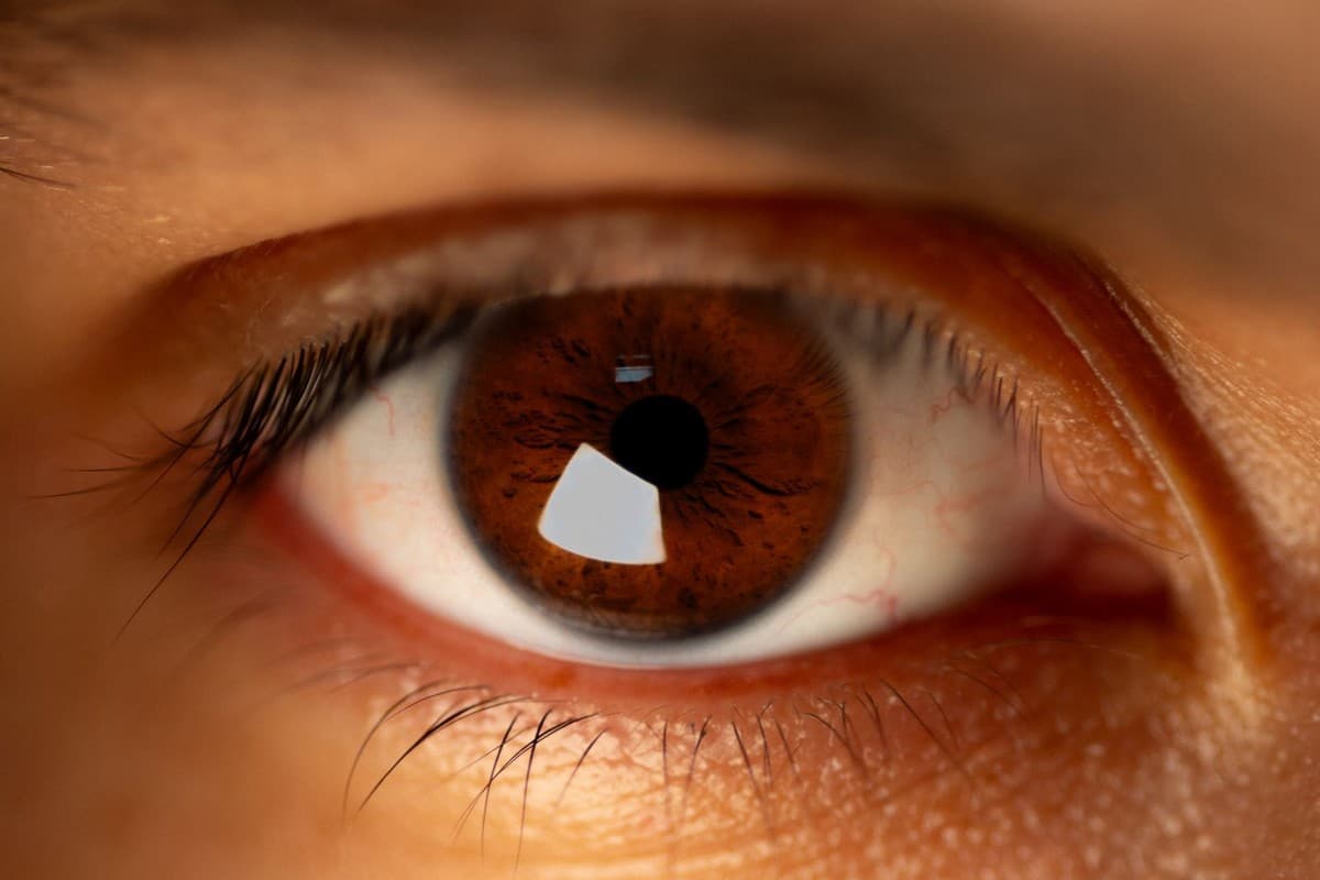
The retina is the layer at the back of the eye that converts light into nerve signals. Problems in this area can affect central or peripheral vision. Macular changes may make reading, recognising faces and seeing straight lines clearly more difficult.
Age-related macular conditions, diabetic retinopathy and retinal vascular occlusions develop for different reasons. Diabetic retinopathy occurs when diabetes affects the retinal blood vessels. Good vision early in a condition does not necessarily mean that the retina is healthy.
An epiretinal membrane is a thin layer of tissue on the macular surface. A macular hole is an opening in the central retinal tissue. Vitreous haemorrhage is bleeding into the clear gel that fills the eye; its cause needs separate investigation.
Symptoms that require assessment without delay
New flashes of light, a sudden increase in floaters or a curtain-like shadow require urgent assessment. These may be associated with a retinal tear or detachment. Symptoms alone cannot establish a diagnosis; an examination of the back of the eye is needed.
Sudden vision loss and new distortion of central vision should not be left waiting either. Retinal problems do not always cause pain. The absence of pain or good vision in the other eye is not enough to dismiss a new change.
Examination of the back of the eye and imaging
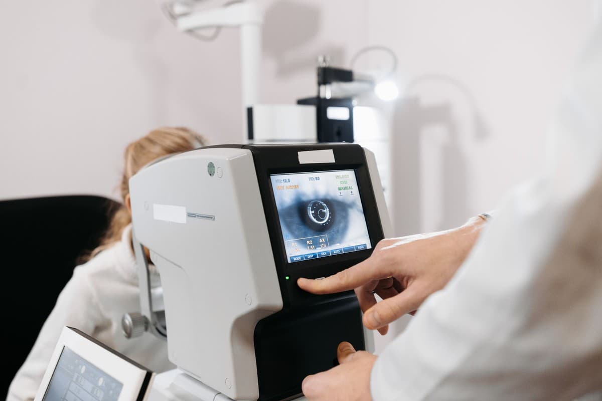
Retinal assessment involves dilating the pupil to examine the back of the eye. An eye doctor in Samsun also assesses the level of vision and when symptoms began. Plan transport home because the drops can cause temporary blurring and light sensitivity.
OCT is a test that produces cross-sectional images of the retinal layers. It can be used to assess swelling, a macular hole or changes in surface tissue. Blood vessel imaging may be added when needed, or ultrasound if the view inside the eye is unclear.
Monitoring, medicines and laser options
Treatment is determined by the type of retinal condition and the degree of threat to vision. Regular monitoring may be sufficient in some cases. Intraocular injections and different laser treatments may be considered for selected vascular or macular conditions.
Retinal laser treatment differs from corneal laser treatment used to correct a glasses prescription. The aim of treatment varies with the condition. A procedure is not used for every macular or retinal problem simply because it works for another retinal condition.
Conditions that require surgery
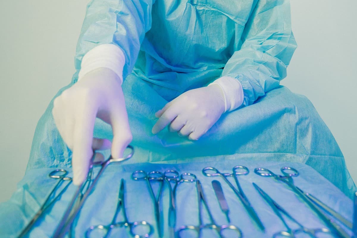
Retinal detachment and some macular or vitreous problems may require surgery. Retinal surgery may include vitrectomy, a procedure that removes the vitreous gel inside the eye. Additional steps, such as removing surface tissue or supporting the retina, are planned when needed.
The decision to operate is not based only on the name of the condition. Its effect on vision, duration, structural findings and the other eye's condition are important. Risks such as infection, pressure changes, the need for another procedure or less visual improvement than expected should be explained.
Specific restrictions after surgery
Flight restrictions are particularly important after surgery that places gas inside the eye. Do not fly until the gas has completely disappeared. The operating surgeon advises on timing; the same number of days does not apply to everyone.
Head positioning may be needed after some operations; the same position is not advised for every patient. If another procedure or anaesthesia is needed, tell the healthcare team that there is gas inside the eye. Do not wait for the scheduled check-up if pain or redness increases or new vision loss occurs.

Long-term eye health and vision support
Some retinal conditions require long-term follow-up. Blood glucose, blood pressure and blood lipid management continue with the relevant doctors. Suitable physical activity and avoiding smoking form part of the general health plan.
Magnifying aids and vision rehabilitation may be considered for lasting vision loss. Rehabilitation supports the use of remaining vision in daily tasks. These measures do not replace treatment of the condition or retinal check-ups.

Retina Care Costs in Samsun in 2026
Cost varies according to investigation and treatment needs. Imaging, medical follow-up, laser treatment and surgery involve different scopes of care. No fixed amount or estimated price is given without assessing the need for repeated check-ups and treatment.
Conclusion
Retina care in Samsun addresses different conditions at the back of the eye through early assessment and individually appropriate follow-up. Timely examination of new symptoms is important. Alongside the goal of preserving vision, treatment selection should consider the nature of the condition, its risks and long-term needs.
Frequently Asked Questions
Can retinal disease progress without pain?
Yes. Some retinal conditions cause no pain and may not produce visual symptoms early on.
Does every floater mean retinal detachment?
No. However, new or suddenly increasing floaters accompanied by flashes or a shadow require examination without delay.
Does OCT replace the entire examination?
No. OCT provides information about specific structures and is interpreted together with a dilated examination of the back of the eye and other findings.
Is flying always prohibited after retinal surgery?
Do not fly while gas remains in the eye. In other circumstances, suitability for travel is discussed according to the operation and follow-up findings.
About this article
Last updated 
Examination and individual assessmentAssoc. Prof. Muhammed Mustafa Kurt, MD
Ophthalmologist · Atakum, Samsun
- Content preparation
- Adapted from information provided for the practice.
- Medical review
- The content update date does not indicate that a medical review has been performed by the physician.
Source: the Word document titled “Retina Care” provided for the practice. No additional medical references were specified in the original text.
This article is for general information and does not replace a medical examination. Diagnosis, suitability for treatment and follow-up are determined individually after an eye examination.