

An eye examination is a series of tests and assessments performed by an ophthalmologist in a specific sequence. During the examination, the patient's visual acuity, intraocular pressure, corneal structure, lens clarity, retina and optic nerve are assessed individually. Specialised instruments such as a slit lamp (biomicroscope), tonometer, ophthalmoscope and autorefractor are used for these assessments. The examination findings are considered together to establish a comprehensive picture of the patient's current eye health.
An eye examination is important not only for investigating the cause of an existing complaint but also for detecting diseases at an early stage, before symptoms develop. Many serious eye diseases, such as glaucoma, diabetic retinopathy and macular degeneration, may not cause noticeable symptoms until they reach advanced stages. Regular eye examinations therefore allow conditions that carry a risk of vision loss to be detected while they are still treatable.
What Is an Eye Examination?
An eye examination is a systematic clinical process that evaluates both the optical function and the anatomical structure of the eye. During the examination, the ophthalmologist measures the patient's level of vision, determines whether a refractive error is present and examines the anterior and posterior segments of the eye in detail. In the anterior segment, the cornea, iris, anterior chamber and lens are assessed, while the retina, macula, optic disc and vitreous are observed in the posterior segment.
This examination is a clinical assessment process that goes far beyond determining a glasses or contact lens prescription. Measuring intraocular pressure, imaging the fundus and planning further investigations when necessary are also integral parts of the examination. The findings provide the basis for various clinical decisions, such as determining the patient's need for treatment, monitoring an existing disease or planning surgery.
Why Is an Eye Examination Important?
Many diseases that threaten eye health can progress without any symptoms in their early stages. Glaucoma is one of the best-known examples, and patients may have no complaints until permanent damage to the optic nerve has occurred. Similarly, conditions such as diabetic retinopathy, retinal vascular occlusions and macular degeneration may progress silently and only become apparent at advanced stages. Regular eye examinations allow these conditions to be detected before they lead to irreversible vision loss.
The importance of an eye examination is not limited to eye diseases. Changes observed in the retinal vessels during a fundus examination may reveal the first signs of systemic diseases such as hypertension and diabetes. Particularly in patients with diabetes, retinal findings provide one of the few clinical windows through which the disease's effects on the body's blood vessels can be directly observed. In this respect, an eye examination is an important screening tool that also contributes to the assessment of overall health.
How Often Should You Have an Eye Examination?
The frequency of eye examinations varies according to a person's age, risk factors and current eye health. Adults with no symptoms or known risk factors are generally advised to have an eye examination at least once every two years between the ages of 20 and 40, and once a year over the age of 40. More frequent check-ups are considered appropriate after the age of 40, as the risk of glaucoma and age-related changes such as presbyopia begin to become more apparent.
People with risk factors such as diabetes, hypertension, a family history of glaucoma or high myopia may need more frequent check-ups, regardless of their age group. An annual fundus examination in patients with diabetes is critical for detecting the development of retinopathy at an early stage. In children, a first comprehensive eye examination at around the age of 3 allows amblyopia and refractive errors to be detected in time during the preschool years.
How Should You Prepare for an Eye Examination?
Considering a few preparations before attending an eye examination helps make the examination more effective. For contact lens wearers, stopping lens use and switching to glasses at least a few days before the examination is important to allow the corneal surface to return to its natural state and to obtain accurate refractive error measurements. Bringing a list of current medications and any reports from previous eye examinations also helps the physician make a more informed assessment.
Pupil dilation may be required during the examination. When dilating eye drops are used, temporary blurred near vision and sensitivity to light may last for several hours. Attending the examination without driving or bringing someone to accompany you may therefore be considered a practical precaution. Avoiding eye makeup before the examination is also recommended, particularly to allow the anterior segment to be assessed properly.
Is an Eye Examination Painful?
An eye examination is generally painless. The instruments used either work without directly touching the eye or, when contact is required, numbing drops are applied to prevent discomfort. Although measuring eye pressure involves light contact with the corneal surface, the anaesthetic drop instilled beforehand means that the patient does not feel this contact. Stages such as slit-lamp examination and fundus examination are performed entirely without contact.
The drops used to dilate the pupils may cause a brief stinging sensation when applied, but this passes within a few seconds. Once the drops take effect, temporary effects such as sensitivity to light and blurred near vision may occur. These effects usually resolve on their own within 4 to 6 hours and do not leave any lasting discomfort.
Stages of an Eye Examination
An eye examination is performed in a specific clinical sequence. First, detailed information is gathered about the patient's complaints, previous eye diseases, systemic diseases, medications and family history. A preliminary refractive error measurement is then taken with an autorefractor, and visual acuity is determined using a Snellen chart. A refraction test establishes the glasses prescription the patient needs for clear vision. Eye pressure is measured with a tonometer to make a preliminary assessment for glaucoma.
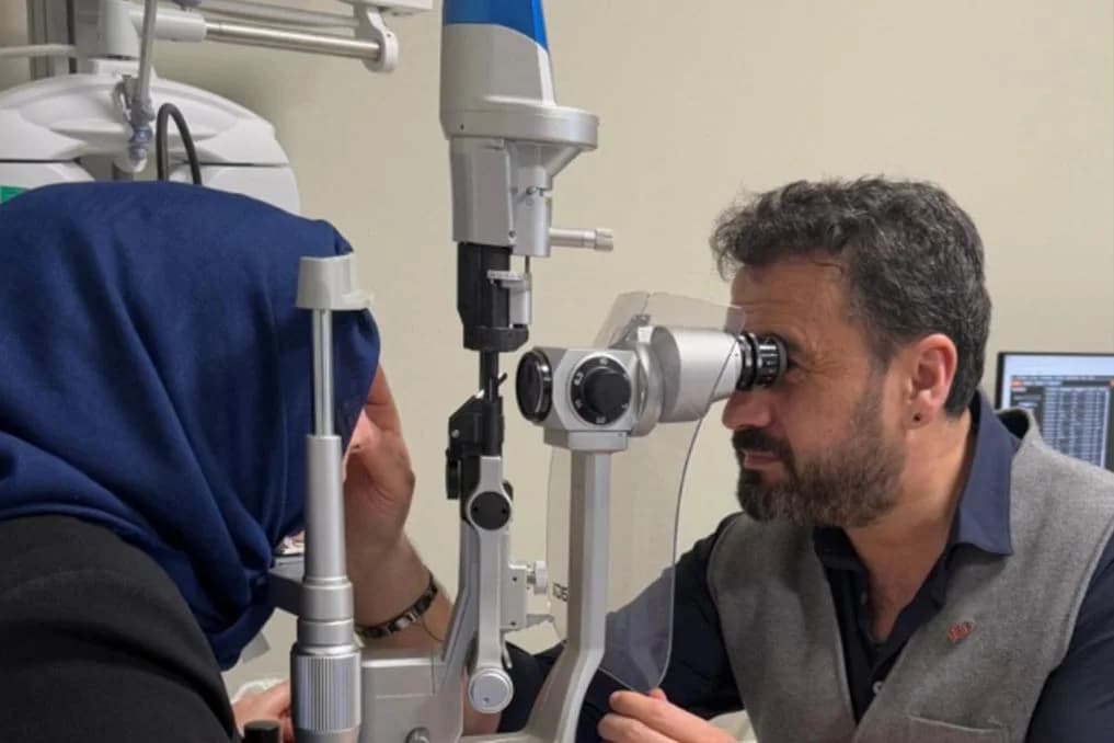
After these basic measurements, the anterior segment of the eye is examined using a biomicroscope (slit lamp). The cornea, iris, anterior chamber depth and lens structure are assessed in detail at this stage. During the fundus examination, the retina, macula, optic disc and retinal vessels are observed with an ophthalmoscope or special lenses attached to the biomicroscope. Where considered necessary, additional tests of eye muscle movement, binocular vision and colour perception may also be included in the examination.
Advanced Investigations Used in Eye Examinations
Advanced investigations are used when findings from a routine eye examination require more detailed assessment. OCT (Optical Coherence Tomography) images the retinal layers at the micron level, playing a critical role in the early diagnosis of conditions such as macular diseases, glaucoma and retinal oedema. Visual field testing (perimetry) maps the patient's peripheral and central vision, making it an indispensable assessment tool for monitoring diseases affecting the optic nerve, particularly glaucoma.
Corneal topography maps the curvature of the corneal surface, providing the data needed to diagnose keratoconus and plan laser treatment. Fundus fluorescein angiography (FFA) uses a dye injected into a vein to image the retinal circulation, allowing vascular abnormalities in conditions such as diabetic retinopathy, retinal vein occlusion and macular degeneration to be examined in detail. The physician determines which advanced investigation is needed according to the examination findings and the patient's clinical presentation.
Diseases Detected Through an Eye Examination
A comprehensive eye examination allows a wide range of eye diseases to be detected. Refractive errors such as myopia, hyperopia, astigmatism and presbyopia are among the most common findings. Cataracts, glaucoma, dry eye syndrome, keratoconus and eyelid diseases are also among the conditions that can be identified during the examination. Examination of the anterior segment of the eye can reveal a large proportion of these diseases.
Fundus examination and advanced investigations can detect diseases affecting the retina and optic nerve. Diabetic retinopathy, macular degeneration, retinal detachment, retinal vascular occlusions, epiretinal membrane and optic neuritis are among the main diseases in this group. An eye examination may also reveal clues to systemic diseases, such as signs of hypertensive retinopathy, the retinal effects of diabetes and even certain rare tumours.
Eye Examination for a Driving Licence
Meeting certain vision standards is a legal requirement for applicants when applying for or renewing a driving licence. An eye examination for a driving licence primarily assesses visual acuity, colour perception and the extent of the visual field. The applicant is expected to have sufficient vision with both eyes or one eye, colour perception that allows traffic signs to be distinguished and the visual field required for safe driving.
The required vision criteria may vary according to the driving licence category. Drivers of heavy goods vehicles and commercial vehicles may be required to meet higher visual acuity standards and have a wider visual field. For people with refractive errors, vision corrected with glasses or contact lenses is taken into account, and a requirement to wear glasses may be recorded on the licence where necessary. People with conditions such as glaucoma, cataracts or retinal disease may need a more detailed assessment during their driving licence examination.
Frequently Asked Questions
How long does an eye examination take?
A standard eye examination takes an average of 15 to 30 minutes. If pupil-dilating drops are used or advanced investigations such as OCT or visual field testing are added, this may extend to between 45 minutes and one hour.
Are pupil-dilating drops used at every examination?
They are not required at every examination. However, pupil dilation may be considered necessary when a detailed fundus assessment is needed, particularly in patients with diabetes, people with suspected retinal disease and patients having a comprehensive examination for the first time.
Can a glasses prescription be determined without an eye examination?
Although an autorefractor provides a preliminary estimate of a glasses prescription, the final prescription can only be determined through a subjective refraction test performed by the physician. Measurements taken at optical stores alone do not provide an adequate assessment and may miss underlying eye diseases.
About this article
Last updated 
Examination and individual assessmentAssoc. Prof. Muhammed Mustafa Kurt, MD
Ophthalmologist · Atakum, Samsun
- Content preparation
- Adapted from an information article on the practice’s existing website.
- Medical review
- The content update date does not indicate that a medical review has been performed by the physician.
Source: the article “How Is an Eye Examination Performed?” on the practice’s existing website. No additional medical references were specified in the original article.
This article is for general information and does not replace a medical examination. Diagnosis, suitability for treatment and follow-up are determined individually after an eye examination.